
The MATE journal publishing platform serves as the central hub for the university’s OJS-based, open access journals.
You can learn more about the MATE Journals service under the "About Us" section on our website.
Below, our journals are presented in alphabetical order.
Journals
-
4D Journal of Landscape Architecture and Garden Art
Journal of Landscape Architecture and Garden Art
ISSN 1787-6613 (Print)
ISSN 2939-886X (Online)The journal 4D is owned by the Institute of Landscape Architecture, Urban Planning and Garden Art at the Hungarian University of Agriculture and Life Sciences. The owner is represented by the Rector of the University.
The journal 4D is owned by the Institute of Landscape Architecture, Urban Planning and Garden Art at the Hungarian University of Agriculture and Life Sciences. The owner is represented by the Rector of the University.
-
Acta Agraria Kaposváriensis
ISSN 1418-1789 (Print)
Founded in 1997 at Kaposvár University. -
Acta Carolus Robertus
ISSN 2498-9312 (Online)
Scientific publications of the Károly Róbert Campus of Szent István University
Founded: 2011 -
-
Acta Scientiarum Socialium
Acta scientiarum socialium : historia, philosophia, sociologia
ISSN 1418-7191 (Print) -
Animal welfare, Ethology and Housing Systems (AWETH)
ISSN 1786-8440 (Online)
Animal Welfare, Etológia és Tartástechnológia journal is an online journal of the Section of Agricultural Sciences of the Hungarian Academy of Sciences. It is published twice a year. The primary language of the journal is Hungarian, but it also publishes scientific articles in Hungarian and English in the fields of animal husbandry, animal breeding, and ethology.
Founded in 2005 at Szent István University.The founder of the scientific journal "Animal Welfare, Etológia és Tartástechnológia" and at the same time its first editor-in-chief (2005-2023) was Dr. János Tőzsér, full professor.
-

MEDIATON OF HUNGARIAN LANGUAGE CULTURE
ISSN 2063-2991 (Online)
Founded: 2012 at Kaposvár University. Restarted in 2019. -
-
COLUMELLA – Journal of Agricultural and Environmental Sciences
ISSN 2064-9479 (Online) ISSN 2064-7816 (Print)
Columella – Journal of Agricultural and Environmental Sciences is an international bi-annual journal that publishes original research articles and review papers in all aspects of agriculture and associated environmental sciences.
Founded: 2014 at Szent István University, Gödöllő -
-
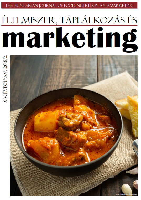
The Hungarian Journal of Food, Nutrition and Marketing
ISSN 2560-2551 (Online)
ISSN 1786-3422 (Printed until 2017-ig) -
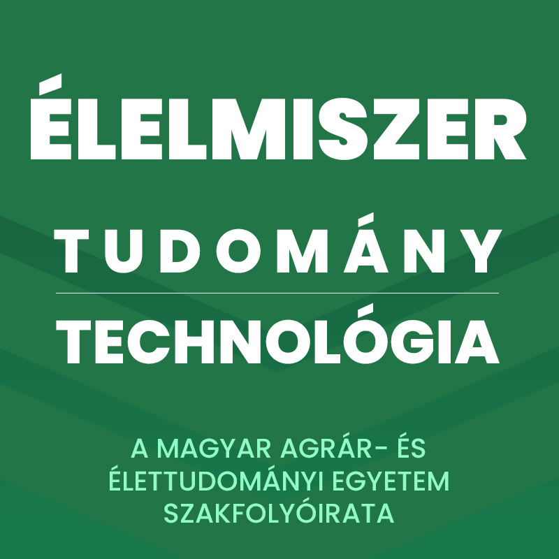
ÉLELMISZER, TUDOMÁNY, TECHNOLÓGIA
ISSN 2061-3954 (Nyomtatott)
A Magyar Agrár- és Élettudományi Egyetem szakfolyóirata.
A folyóirat célja a kutatók és a szakemberek folyamatos tájékoztatása az élelmiszerszektor teljes vertikumában fellelhető legújabb, legfrissebb eredményekről, eseményekről, fejlesztésekről és beruházásokról. -
GEORGIKON FOR AGRICULTURE
ISSN 0239-1260 (print)
A multidisciplinary journal in agricultural sciences. The Journal Georgikon for Agriculture (briefly: G.Agric) is published twice a year by Hungarian University of Agriculture and Life Sciences, Georgicon Campus. Articles of original research findings in all fields of agriculture and related topics are published in the Journal subsequent to critical revieww and approval by the Editorial Board. -
-

Training and Practice : Journal of Educational Sciences
ISSN 2064-4027 (online)
-
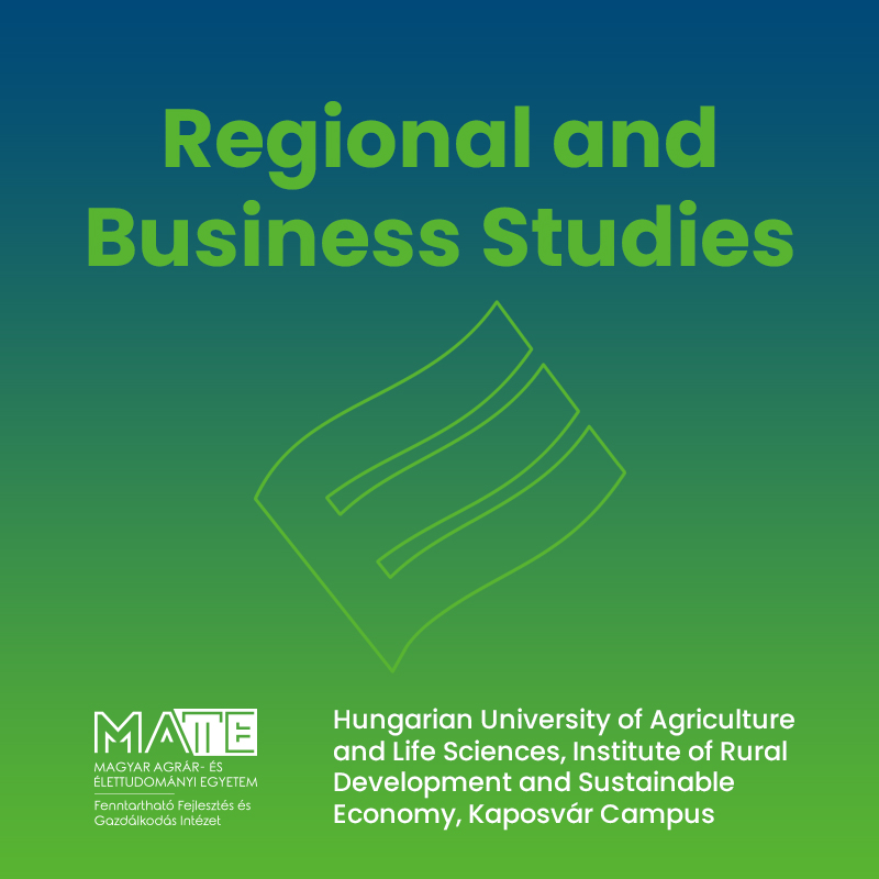
REGIONAL AND BUSINESS STUDIES
ISSN 2732-2726 (Online) ISSN 2061-2311 (Print until 2020)
Regional and Business Studies is an international, transdisciplinary scientific journal published recently twice a year and contains original scientific reports, research results, critical résumés, conference reviews, and letters to the editors. The topic of the journal includes the important fields of business, rural and regional development, and social sciences.
Founded: 2009 at Kaposvár University Faculty of Economic Science. -

-

Technical Languages and Translation
ISSN 1587-4389 (Nyomtatott)
Tanulmányok a Magyar Agrár- és Élettudományi Egyetem szaknyelvi és szakfordítással kapcsolatos kutatásainak eredményeiről.
Alapítva a Szent István Egyetem Gazdaság- és Társadalomtudományi Kar Alkalmazott Nyelvészeti Tanszékén. -
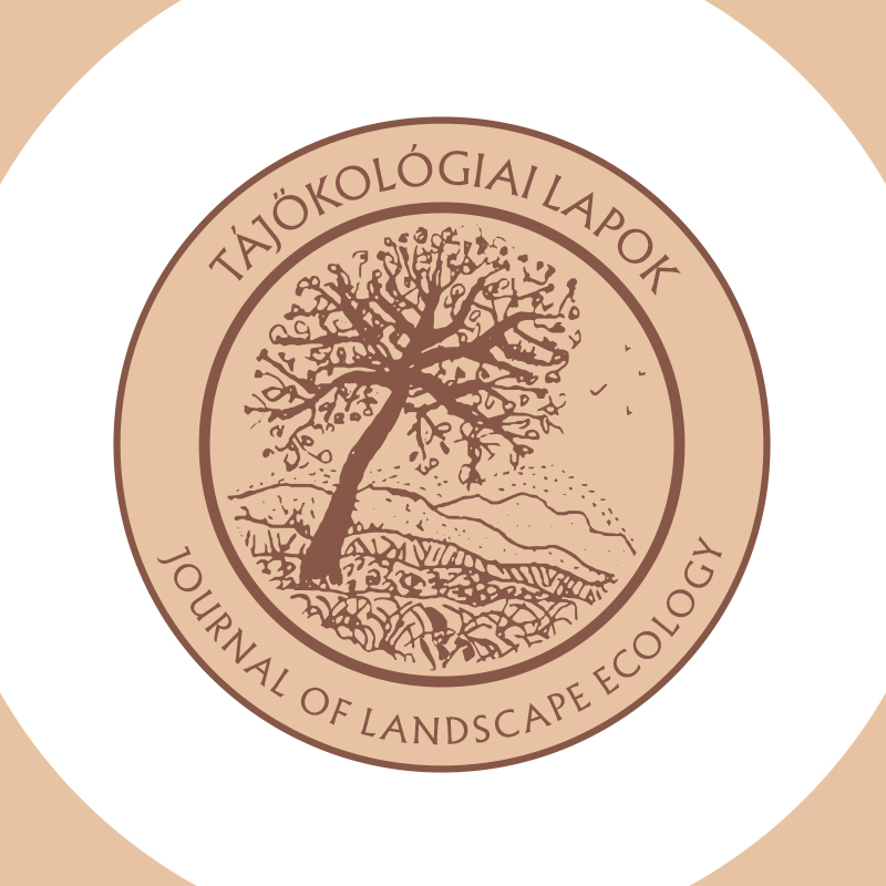
JOURNAL OF LANDSCAPE ECOLOGY
ISSN 1589-4673 (Print)
In our present world new scientific areas are arising. Basic researches are developing, areas handling our environment more as units are under intensive development. In these works the role of protection and knowing of the landscape has increased significantly. Our journal started in 2003 wishes to participate in the development of landscape ecology as a synthesizing science and disciplines directly connecting to it. Our journal provides opportunity for scientists to publish new results, to write publications in Hungarian and in other world languages, too. We are waiting for short communications that help scientists in scientific orientation.
